Warning: mkdir(): Permission denied in /home/virtual/lib/view_data.php on line 81 Warning: fopen(/home/virtual/neurointervention/journal/upload/ip_log/ip_log_2024-04.txt): failed to open stream: No such file or directory in /home/virtual/lib/view_data.php on line 83 Warning: fwrite() expects parameter 1 to be resource, boolean given in /home/virtual/lib/view_data.php on line 84 Fully Reversible Contrast-Induced Encephalopathy Mimicking Stroke after Flow Diverter Treatment of Carotid Cave Aneurysm
Neurointervention Search

CLOSE


Neurointervention > Volume 18(1); 2023 > Article
Diamandis, Swiatek, and Behme: Fully Reversible Contrast-Induced Encephalopathy Mimicking Stroke after Flow Diverter Treatment of Carotid Cave Aneurysm

Abstract

Contrast-induced encephalopathy (CIE) is a rare complication of coronary and neurointerventional procedures. The condition is believed to arise from endothelial damage secondary to exposure to iodinated contrast media. A wide spectrum of clinical manifestations has been reported including seizures, cortical blindness, and focal neurological deficits. This report details the case of fully reversible CIE mimicking severe anterior circulation stroke in a 55-year-old female following elective endovascular treatment with a flow diverter of a carotid cave aneurysm. The patient was managed conservatively with intravenous hydration and steroids and showed an excellent prognosis with supportive management.

INTRODUCTION

Contrast-induced encephalopathy (CIE) is a rare condition complicating about 1–4% of neuroendovascular procedures [1,2]. It usually presents with rapidly deteriorating neurological deficits in the hours following angiographic procedures involving intra-arterial administration of iodinated contrast media [3]. The most commonly reported symptoms are headaches, seizures, and focal neurological deficits including transient global amnesia, cortical blindness, ophthalmoplegia, and hemiparesis [4]. Although mostly transient and reversible, the symptoms may be persistent with permanent sequelae and adverse clinical outcomes [5].
Herein, we report a case of CIE following flow diverter treatment that mimicked a severe anterior circulation stroke.

CASE REPORT

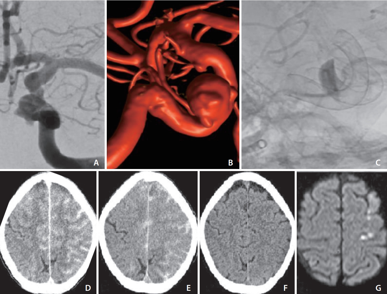
A 55-year-old Caucasian female without any clinical premorbidities was admitted to our hospital to undergo elective endovascular treatment of a carotid cave aneurysm of the left internal carotid artery (ICA), measuring approximately 9 mm in its biggest diameter (Fig. 1A, B).
The patient was under dual antiplatelet therapy with aspirin (100 mg/d) and clopidogrel (75 mg/d) following the treatment of a paraophthalmic aneurysm of the right ICA with a flow diverter (Derivo 2 Heal; Acandis, Pforzheim, Germany) 6 weeks earlier. A total of 70 mL of non-diluted contrast (Iohexol, Accupaque™ 300 mg/mL; GE Healthcare, Chicago, IL, USA) was used without any complications.
Induction in general anesthesia was performed, and standard monitoring was obtained. A triaxial system was introduced through a femoral access using a long sheath (Cerebase DA Guide Sheath; Cerenovus, New Brunswick, NJ, USA), an intermediate catheter (AXS Catalyst6; Stryker, Kalamazoo, MI, USA), and a 0.027 inch microcatheter (Neuroslider; Acandis). A 5x15 mm flow-diverter (Derivo 2; Acandis) was implanted with optimal wall apposition and a clear intraaneurysmal contrast stagnation (Fig. 1C).
Multiple angiographic runs and 3D rotational angiograms (3DRAs) were performed during the procedure, using a total of 125 mL non-diluted contrast (Iohexol, Accupaque™ 300 mg/mL; GE Healthcare).
Right after extubation, the patient presented with hemiparesis of the right upper and lower extremities with hemineglect towards the paretic side.
Approximately 45 minutes after the procedure, the patient became progressively aphasic, which rapidly developed into a complete receptive and expressive aphasia.
Non-contrast computed tomography (NCCT), computed tomography (CT)-angiography, and CT-perfusion were immediately performed to rule out stroke. The NCCT showed diffuse gyral hyperdensities mimicking sulcal subarachnoid hemorrhage in the entire left ICA territory with similar changes in the right anterior cerebral artery territory. Hyperdensity of the left basal ganglia was noticed (Fig. 1D). Subsequently, the patient was admitted to the neurosurgical intensive care unit where supportive therapy with intravenous (IV) fluids and corticosteroids (prednisolone 8 mg/d) was administered. No epileptic activity was observed. Four hours later, a repeat NCCT was obtained which showed a mild regression of the aforementioned changes (Fig. 1E).
Twenty hours after the procedure, the patient regained motor function in the lower extremity with persistent global aphasia and paresis of the right upper extremity.
A repeat NCCT performed 30 hours after the procedure showed complete disappearance of the earlier changes. No ischemic findings were noticed (Fig. 1F).
Over the course of the following 3 days, the symptoms gradually disappeared with the regaining of motor function of the right upper extremity.
The patient was discharged to the general ward on postprocedural day 4. On the fifth postprocedural day an magnetic resonance imaging (MRI) was performed, which showed a few punctate cortical diffusion-weighted imaging (DWI) lesions in the precentral, postcentral, and middle frontal Gyrus (Fig. 1G). No susceptibility foci were identified on T2*.
The patient was discharged from our hospital on the seventh postprocedural day. Her neurological examination at the time of discharge was unremarkable, except for dysgraphia. A neurological rehabilitation program was completed by the patient, which resulted in complete neurologic recovery at 3 weeks after discharge.

DISCUSSION

Pathophysiology and Risk Factors

CIE is a rare complication of diagnostic and interventional angiography procedures involving coronary and cerebral arteries. The pathophysiologic mechanism appears to be related to the disruption of the blood brain barrier after application of iodinated contrast media [6]. Non-ionic and low-osmolar contrast agents have also been reported to cause CIE [7]. These have been associated with a higher incidence of CIE of about 4% [4,8]. Iohexol, a low-osmolar non-ionic monomer, was used in this case.
It has been suggested that higher doses and lower temperatures of contrast media, kidney disorders, arterial hypertension, and acute cerebral infarction are risk factors for CIE [9]. Higher contrast dose and prior stroke were corroborated as risk factors in a recent meta-analysis [10]. However, lower doses of contrast agent have also been associated with CIE [11].
The doses reported in the literature varied widely depending on procedure type with volumes as low as 24 mL in a cerebral angiography case [6] and as high as 360 mL in a spinal angiography case [12]. Our patient received a total volume of contrast of 125 mL.

Clinical Manifestation

The hyperacute onset of symptoms after cerebral or coronary angiography and the reversibility of clinical findings are hallmarks in the diagnosis of CIE [13]. In a literature review on CIE following neurointerventional procedures, the most commonly reported symptom onset time was either immediately or within a few hours after angiography [14]. A prodromal onset pattern with gradual progression of symptoms over 17 hours has been reported in a case of a fatal irreversible CIE [15].
Late-onset imaging-confirmed encephalopathy cases with intervals of 1 week to 1 month following cerebral angiography have been attributed to posterior reversible encephalopathy syndrome and an inflammatory response to coating materials [16–18].
The clinical syndrome is usually reversible within 24–72 hours. Longer duration and irreversible or fatal outcomes, however, have also been reported [15,19].
A wide range of neurological deficits can occur depending on the arterial territory involved. Transient cortical blindness (TCB) is the most commonly reported manifestation of CIE related to coronary angiography. In cerebral angiography, TCB is mainly related to vertebral artery angiography [20].

Imaging Features

The main CT features of CIE are cortical swelling with poorly localized enhancement and hyperdensity of subarachnoid space [21] that may mimic subarachnoid hemorrhage. MRI usually shows T2/Flair hyperintensities in the involved territories with foci of diffusion-restriction in DWI [3]. Reversibility of these features in the subacute stage is the key to the diagnosis of CIE. This was also the case with our patient.

Literature Review

Different rates of CIE have been reported depending on procedure type, varying from 0.66% in coil embolization [22] to 1.7% in mechanical thrombectomy [1]. An incidence of 2.9% has been reported in a case series on TCB following coil embolization of posterior circulation aneurysms [23], suggesting a higher incidence of CIE in posterior circulation interventions. In comparison, CIE appears to be much less common in coronary angiography with reported incidences of 0.05–0.4% [24]. This could be explained by the frequent injections of large volumes of contrast directly into a single cerebral vessel in neurointervention [7].

Management and Prevention

Despite the lack of clinical evidence on the management of CIE, early supportive therapy with aggressive IV hydration and administration of steroids is the most commonly reported management strategy [25]. Our patient was managed conservatively with IV fluids and was monitored for epileptic activity in the early phase. No prophylactic anticonvulsant was administered.
Awareness of CIE and its typical clinical and radiological findings among interventionalists is essential for early recognition, correct diagnosis, and management. In patients with a history of CIE, avoidance of subsequent administrations of contrast media intra-arterially might be warranted. Stringent use of contrast in neurointerventional procedures avoiding multiple 3DRAs and distal access catheter injection seems advisable from our point of view.

Notes

Fund

None.

Ethics Statement

Institutional Review Board (IRB) approval for case reports is not required at our institution. We obtained individual written informed consents for the procedure and for publication.

Conflicts of Interest

The authors have no conflicts to disclose.

Author Contributions

Concept and design: AVN and EAB. Analysis and interpretation: AVN and EAB. Data collection: AVN and EAB. Writing the article: AVN and EAB. Critical revision of the article: AVN and EAB. Final approval of the article: AVN and EAB.

Fig. 1.
(A) Anteroposterior angiographic run showing carotid cave aneurysm of left internal carotid artery (ICA). (B) 3D rotational angiogram of the same aneurysm. (C) Unsubstracted image after flow diverter implantation showing intraaneurysmal contrast stagnation and also showing the superimposed flow diverter of the right ICA implanted 6 weeks earlier. (D) Post-procedural computed tomography (CT) showing sulcal hyperdensities mimicking subarachnoid hemorrhage. (E, F) Repeat non-contrast CT at 4 hours and 30 hours, respectively, showing gradual regression of previous findings and ruling out ischemic changes. (G) Magnetic resonance imaging on postprocedural day 5 showing a few punctate diffusion-weighted imaging hits.
neuroint-2022-00353f1.jpg

REFERENCES

1. Chu YT, Lee KP, Chen CH, Sung PS, Lin YH, Lee CW, et al. Contrast-induced encephalopathy after endovascular thrombectomy for acute ischemic stroke. Stroke 2020;51:3756-3759.
crossref pmid
2. Potsi S, Chourmouzi D, Moumtzouoglou A, Nikiforaki A, Gkouvas K, Drevelegas A. Transient contrast encephalopathy after carotid angiography mimicking diffuse subarachnoid haemorrhage. Neurol Sci 2012;33:445-448.
crossref pmid pdf
3. Yu J, Dangas G. Commentary: new insights into the risk factors of contrast-induced encephalopathy. J Endovasc Ther 2011;18:545-546.
crossref pmid
4. Leong S, Fanning NF. Persistent neurological deficit from iodinated contrast encephalopathy following intracranial aneurysm coiling. A case report and review of the literature. Interv Neuroradiol 2012;18:33-41.
crossref pmid pmc pdf
5. Cristaldi PMF, Polistena A, Patassini M, de Laurentis C, Giussani C, Remida P. Contrast-induced encephalopathy and permanent neurological deficit: a case report and literature review. Surg Neurol Int 2021;12:273
crossref pmid pmc
6. Lantos G. Cortical blindness due to osmotic disruption of the blood-brain barrier by angiographic contrast material: CT and MRI studies. Neurology 1989;39:567-571.
crossref pmid
7. Uchiyama Y, Abe T, Hirohata M, Tanaka N, Kojima K, Nishimura H, et al. Blood brain-barrier disruption of nonionic iodinated contrast medium following coil embolization of a ruptured intracerebral aneurysm. AJNR Am J Neuroradiol 2004;25:1783-1786 Erratum in: AJNR Am J Neuroradiol 2005;26:203

8. Liu MR, Jiang H, Li XL, Yang P. Case report and literature review on low-osmolar, non-ionic iodine-based contrast-induced encephalopathy. Clin Interv Aging 2020;15:2277-2289.
pmid pmc
9. Iwata T, Mori T, Tajiri H, Miyazaki Y, Nakazaki M. Repeated injection of contrast medium inducing dysfunction of the bloodbrain barrier: case report. Neurol Med Chir (Tokyo) 2013;53:34-36.
pmid
10. Vazquez S, Graifman G, Spirollari E, Ng C, Uddin A, Feldstein E, et al. Incidence and risk factors for acute transient contrast-induced neurologic deficit: a systematic review with meta-analysis. Stroke Vasc Interv Neurol 2022;2:e000142.
crossref
11. Kamimura T, Nakamori M, Imamura E, Hayashi Y, Matsushima H, Mizoue T, et al. Low-dose contrast-induced encephalopathy during diagnostic cerebral angiography. Intern Med 2021;60:629-633.
crossref pmid pmc
12. Eckel TS, Breiter SN, Monsein LH. Subarachnoid contrast enhancement after spinal angiography mimicking diffuse subarachnoid hemorrhage. AJR Am J Roentgenol 1998;170:503-505.
crossref pmid
13. Spina R, Simon N, Markus R, Muller DW, Kathir K. Contrast-induced encephalopathy following cardiac catheterization. Catheter Cardiovasc Interv 2017;90:257-268.
crossref pmid pdf
14. Nagamine Y, Hayashi T, Kakehi Y, Yamane F, Ishihara S, Uchino A, et al. Contrast-induced encephalopathy after coil embolization of an unruptured internal carotid artery aneurysm. Intern Med 2014;53:2133-2138.
crossref pmid
15. Zhao W, Zhang J, Song Y, Sun L, Zheng M, Yin H, et al. Irreversible fatal contrast-induced encephalopathy: a case report. BMC Neurol 2019;19:46
crossref pmid pmc pdf
16. Jang HW, Lee HJ. Posterior reversible leukoencephalopathy due to “triple H“ therapy. J Clin Neurosci 2010;17:1059-1061.
crossref pmid
17. Giraldo EA, Fugate JE, Rabinstein AA, Lanzino G, Wijdicks EF. Posterior reversible encephalopathy syndrome associated with hemodynamic augmentation in aneurysmal subarachnoid hemorrhage. Neurocrit Care 2011;14:427-432.
crossref pmid pdf
18. Skolarus LE, Gemmete JJ, Braley T, Morgenstern LB, Pandey A. Abnormal white matter changes after cerebral aneurysm treatment with polyglycolic-polylactic acid coils. World Neurosurg 2010;74:640-644.
crossref pmid
19. Allison C, Sharma V, Park J, Schirmer CM, Zand R. Contrast-induced encephalopathy after cerebral angiogram: a case series and review of literature. Case Rep Neurol 2021;13:405-413.
crossref pmid pmc pdf
20. Lo LW, Chan HF, Ma KF, Cheng LF, Chan TK. Transient cortical blindness following vertebral angiography: a case report. Neurointervention 2015;10:39-42.
crossref pmid pmc
21. Meijer FJA, Steens SCA, Tuladhar AM, van Dijk ED, Boogaarts HD. Contrast-induced encephalopathy-neuroimaging findings and clinical relevance. Neuroradiology 2022;64:1265-1268.
crossref pmid pmc pdf
22. Matsubara N, Izumi T, Miyachi S, Ota K, Wakabayashi T. Contrast-induced encephalopathy following embolization of intracranial aneurysms in hemodialysis patients. Neurol Med Chir (Tokyo) 2017;57:641-648.
crossref pmid pmc
23. Niimi Y, Kupersmith MJ, Ahmad S, Song J, Berenstein A. Cortical blindness, transient and otherwise, associated with detachable coil embolization of intracranial aneurysms. AJNR Am J Neuroradiol 2008;29:603-607.
crossref pmid pmc
24. Andone S, Balasa R, Barcutean L, Bajko Z, Ion V, Motataianu A, et al. Contrast medium-induced encephalopathy after coronary angiography- case report. J Crit Care Med (Targu Mures) 2021;7:145-149.
crossref pmid pmc
25. Shinoda J, Ajimi Y, Yamada M, Onozuka S. Cortical blindness during coil embolization of an unruptured intracranial aneurysm--case report. Neurol Med Chir (Tokyo) 2004;44:416-419.
crossref pmid
TOOLS
Share :
Facebook Twitter Linked In Google+ Line it
METRICS Graph View
  • 1 Web of Science
  • 2 Crossref
  •    
  • 2,814 View
  • 121 Download


ABOUT
ARTICLE CATEGORY

Browse all articles >

BROWSE ARTICLES
AUTHOR INFORMATION
Editorial Office
#705-2C, 9 Gangnam-ro, Giheung-gu, Yongin-si, Gyeonggi-do 16977, KOREA
Tel: +82-31-994-4382    Fax: +82-2-2275-9198    E-mail: editor@neurointervention.org                

Copyright © 2024 by Korean Society of Interventional Neuroradiology.

Developed in M2PI

Close layer
prev next