INTRODUCTION
Mechanical thrombectomy is the well-accepted standard of care for acute large vessel occlusions in the internal carotid artery (ICA) and in the M1 segment of the middle cerebral artery (MCA) [1]. The safety and efficacy of thrombectomy in more distal intracranial branches are on the other hand less clear. Recent observational studies showed promising results for distal M2 and M3 occlusions treated with stent retrieval thrombectomy [2]. Occluded distal branches that supply areas of functionally significant parenchyma could potentially benefit from endovascular thrombectomy in selected cases, but, feasibility of this technique is limited by the distal thrombus location and by the tortuosity and potential fragility of these small vessels, hence harbouring a presumed higher risk of complications.
MATERIALS AND METHODS
Endovascular technique
After confirmation of the vessel occlusion at the M2/3 segment of MCA by computed tomography (CT) and selective catheter angiography (Fig. 1), a Flowgate balloon guiding catheter (Stryker, Kalamazoo, MI, USA) was placed in the distal cervical ICA and a Trevo Pro 14 (Stryker) microcatheter was then navigated past the occlusion over a Synchro 14 (Stryker) micro guidewire. In light of the small calibre and tortuosity of the occluded vessel, a small Trevo XP ProVue 3├Ś20 mm stent retriever (Stryker) was only partially deployed within the thrombus, leaving half of the stent length sheathed inside the microcatheter (Fig. 2A, B). The stent was kept open for 5 minutes for thrombus engagement and then retrieved under proximal flow reversal at the balloon guiding catheter, resulting in revascularization of the affected MCA territory (Fig. 2C, D).
Illustrative cases
Case 1
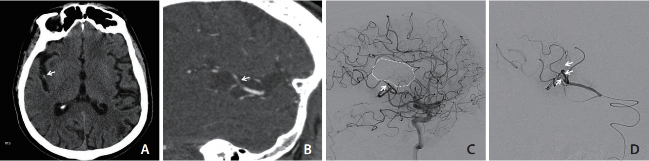
A 68-year-old male patient presented with acute left hemiplegia 2 hours before admission. Due to the left leg weakness, the patient fell and suffered a subtrochanteric fracture of his left femur. The initial National Institute of Health Stroke Scale (NIHSS) was 8. There was no intracranial haemorrhage and the Alberta Stroke Program Early CT Score was 10. A CT-angiogram revealed a 7 mm-long filling defect in the superior division (M2 trunk) of right MCA, with a modified collateral score of 3 (Fig. 1A, B) [3]. Despite the early presentation, intravenous thrombolysis (IV) was contraindicated due to the long bone fracture. The patient was then referred for endovascular thrombectomy.
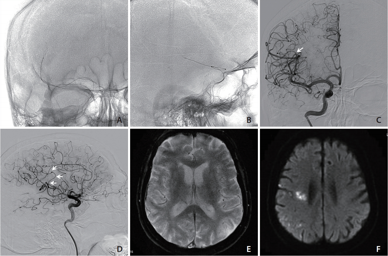
An initial ICA angiogram demonstrated that the M2 thrombus had migrated distally into the origin of right precentral artery (M3), a vessel measuring 1.6 mm in diameter (Fig 1C, D). Considering the functional eloquence of the motor cortex supplied by this occluded branch, it was decided to attempt revascularization of this branch. Using a 3 mm stent-retriever and the semi-deployment technique, modified treatment in cerebral infarction (mTICI) 3 revascularization of the affected MCA territory was achieved (Fig. 2C, D). The groin puncture-to-reperfusion time was within 35 minutes.
The NIHSS improved from 8 to 2 in the first 24 hours after treatment, with resolution of the left hemiparesis. The patient underwent uneventful fixation of the left femoral fracture and was discharged to home within 1 week. A follow-up magnetic resonance imaging performed 3 days after thrombectomy showed patent MCA branches, a few scattered small foci of restricted diffusion in the right hemisphere suggestive of small distal emboli without evidence of large regional infarctions, and no hemorrhagic complications (Fig. 2E, F). The modified Rankin scale at discharge was 1.
Case 2
A 71-year-old man with left carotid bifurcation stenosis was admitted for acute right hemiplegia. The initial CT angiogram and subsequent catheter angiogram confirmed occlusion of the inferior M2 territory, obstructing the precentral artery (Fig. 3A, B). mTICI 3 revascularization was achieved with the semi-deployment of a Trevo XP ProVue 3├Ś20 mm stent retriever (Fig. 3C, D). The left cervical carotid artery was subsequently treated with stenting and angioplasty (Fig. 3E, F). The modified Rankin scale at discharge was 1.
DISCUSSION
Depending on the ŌĆ£eloquenceŌĆØ of the supplied territory, small cerebral vessel occlusions can lead to severe neurological deficits. While IV with tissue plasminogen activator (tPA) remains the standard of care, its use is frequently limited by the short therapeutic time window and by multiple contraindications, such as the long bone fracture in our first patient.
Endovascular mechanical thrombectomy provides rapid recanalization and improves the neurological outcome in large vessel occlusions of the ICA and M1 segment of MCA [4]. This technique has been increasingly applied to M2 and M3 occlusions, but its benefits over medical treatment with IV tPA and its safety profile have not been clearly demonstrated. In particular, the risk of vessel perforation and of haemorrhagic complications remains a concern [5].
Haussen et al. [5] reported four cases of M3 occlusions treated with thrombectomy with a 3 mm Trevo stentriever. Two of these patients suffered from intracranial haemorrhage after mechanical thrombectomy that led to poor outcomes. This relatively high rate of haemorrhagic complications may in part be due to the radial and tractional force exerted by the stent struts on these small and often tortuous vessels during retrieval of the device, resulting in perforator or venous avulsion or vessel laceration.
Using in-vitro models, Machi et al. [6], demonstrated that the radial pressure (RN) exerted by stent retrievers on the vessel wall is inversely proportional to the length of the deployed stent. This was expressed by the formula RN = F / (2ŽĆrL╬╝), where r is the internal radius of the blood vessel, L is the length of the deployed stent and ╬╝ is the friction coef’¼ücient [6]. We therefore elected to deploy half of the working length of the ŌĆ£Baby TrevoŌĆØ stentriever in the precentral artery with just adequate length to engage the thrombus, avoiding unnecessary length of stent open across the sharp angles of the vessel. The thrombus was successfully removed with one pass and no hemorrhagic complications were encountered in both cases. A similar concept was first described by Imahori et al. [7] in 14 cases of M1 or M2 occlusion using larger stent retrievers if the blood clot is short.
Aspiration thrombectomy with smaller reperfusion catheters is an alternative therapy for distal occlusions. Premat et al. [8] reported good navigability of the 3 max reperfusion catheter (Penumbra Inc., Alameda, CA, USA) in M3 branches and treated six M3 occlusions. However, the reperfusion rate by aspiration alone seems to be lower than stent retrievers for small vessel occlusions, especially in those just distal to an angulated vessel segment [9]. Stent retrieval technique also appears to have a higher first pass reperfusion rate as a standalone first-line approach to ICA/M1/M2 occlusions [10]. The current case report suggests that semi-deployment of small stent retriever may be a safe and effective treatment strategy for distal occlusions.
CONCLUSION
For acute distal occlusions in small but functionally eloquent vessels such as M2/3 branches supplying the motor cortex, mechanical thrombectomy with small calibre stentrievers is reasonable and feasible. Semi-deployment of the stent retriever in small and tortuous vessels to reduce radial and tractional forces on the vascular wall can potentially avoid hemorrhagic complication associated with these distal thrombectomies.