INTRODUCTION
Transarterial embolization with Onyx (Medtronic) is currently an established method of treatment for dural arteriovenous fistulas (DAVFs) [1,2]. Onyx is cohesive and nonthrombogenic, which allows for better packing of lesions and makes venous occlusion less likely than n-butyl cyanoacrylate. Onyx occasionally passes to the draining vein during the packing of the shunt portions, but the cast usually slowly moves forward and hardens at the entrance of the draining vein side while sticking to the draining veinŌĆÖs wall. However, we encountered a unique finding of intravenous Onyx migration during transarterial embolization of a transverse sigmoid sinus DAVF. A hairball-like structure suddenly appeared in the jugular vein during Onyx injection. It was removed transvenously and confirmed to be an Onyx thread on pathological examination. This brief report aimed to determine the mechanism underlying this abnormal phenomenon in an experimental simulation.
CLINICAL CASE AND EXPERIMENTAL VALIDATION
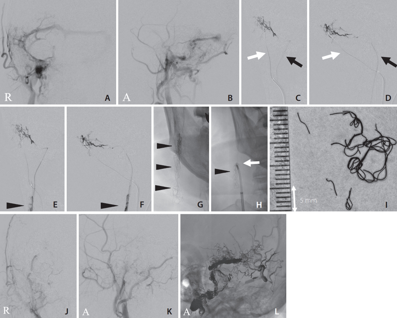
A 66-year-old man presenting with pulsatile tinnitus was diagnosed with a right jugular bulb and transverse sigmoid sinus DAVF and was referred to our hospital for endovascular treatment (Fig. 1A, B). Under general anesthesia, 6-Fr long sheaths were inserted into the right femoral artery and vein. A 6-Fr guiding catheter was navigated into the right jugular vein and external carotid artery. First, we performed a transvenous embolization of the fistula in the jugular bulb. A Headway 17 microcatheter (Terumo) was navigated through a shunting pouch around the right anterior condylar confluence. The detachable coils were deployed in a shunting pouch. Then, a transarterial embolization of the right external carotid artery was performed. The feeders of the right ascending pharyngeal, right posterior auricular, and right middle meningeal arteries were embolized using Onyx-18 (Medtronic). Finally, a Scepter balloon catheter 4├Ś7 mm (Terumo) was navigated into the right occipital artery, and Onyx was administered. We inflated a Shouryu balloon catheter 7├Ś7 mm (Kaneka) inside the right transverse sigmoid sinus to avoid Onyx migration into the venous side during injection (Fig. 1C, D). An abnormal thread-like black line connected to the injection site was observed on the digital subtraction angiography monitor during Onyx injection from the Scepter balloon catheter (Terumo). The origin of this thread was proximal to the inflated balloon for sinus protection. It accumulated in the jugular bulb as a hairball-like mass (Fig. 1E, F). The injection was stopped, and the Scepter catheter was removed. The foreign body in the jugular vein was retrieved using a goose-neck snare 10 mm (Medtronic) (Fig. 1G, H), and a black thread-like mass was identified (Fig. 1I). The pathological result of the foreign body was revealed to be Onyx. The transverse sigmoid sinus DAVF was treated with transvenous sinus packing using coils (Fig. 1JŌĆōL). Almost all the shunts disappeared, as well as the patientŌĆÖs tinnitus, immediately after the operation.
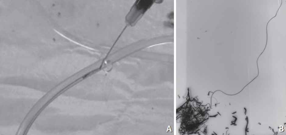
We planned an experimental model to determine the mechanism underlying this abnormal phenomenon. We hypothesized that Onyx precipitates in a thread-like shape when passing through an extremely narrow hole. Therefore, Onyx was extruded from a syringe using a 27-gauge needle into a silicone tube with flowing water. In this experiment, we changed the flow speed of the water and observed the hardening behavior of Onyx (Fig. 2). The extruded Onyx quickly hardened at the needle tip and formed a round mass under very slow flow. Onyx was blown away as small scattering pieces under high-speed flow. Onyx threads were successfully formed only under the adequate continuous slow flow condition (Supplementary Video 1).
DISCUSSION
Transarterial Onyx embolization for DAVFs is a safe and effective therapeutic alternative [1,2]. However, migration of Onyx to the venous side through the shunt point impairs normal venous drainage and increases the risk of pulmonary embolism. Wang et al. [3] reported a case of cardiac Onyx migration that required thoracic surgery. They also reported a case of deep cerebral venous occlusion that led to deterioration or death. Therefore, we usually use a large protective balloon in the affected sinus to avoid such venous migration and preserve normal sinus blood flow.
Transarterial balloon-assisted embolization combined with transvenous sinus balloon protection using a transvenous balloon is reportedly a useful adjunctive technique for the treatment of DAVFs [4]. Using this method, greater penetration into the shunting points of Onyx and less migration into the venous side can be expected compared with other conventional methods. However, the shape of the cross-section of the sinus is not round, and many trabeculations exist in the cavity. Therefore, complete balloon attachment to the sinus wall is difficult, and the balloon does not always cover all shunt points for DAVF with multiple shunt points, similar to our case. We used a relatively short balloon because a long, super-compliant balloon (e.g., 80 mm in length) is not approved for use in Japan. Therefore, migration of Onyx to the venous side occurred.
Onyx penetrating the venous side usually forms a claylike mass at the exit of the shunt point. However, in this case, Onyx hardened into a thread-like shape and continuously jetted out without breaking. If this mass had migrated into the pulmonary artery, a serious complication would have occurred; however, the Onyx threads remained in the jugular vein in 1 lump, forming a hairball. Therefore, it was easily retrieved using a goose-neck snare.
Our study demonstrated that the extruded Onyx quickly hardened at the needle tip and formed a round mass under very slow flow, whereas Onyx was blown away as small scattering pieces under high-speed flow. Onyx threads were successfully formed only under an adequate continuous slow flow condition, similar to our case. These results indicate that transarterially injected Onyx penetrated through a tiny channel connected to the sinus wall and precipitated continuously by applying constant pressure in a less pulsatile and stable venous flow in the sinus. The size of the shunt area varies, as previously reported, with cases ranging from micro-pinholes to >3 mm in diameter [5], but accidents during the transarterial injection of Onyx are caused by tiny channels in the perisinus network directly connected to the sinus. Onyx threads may be overlooked because they are thin and sway, and the operator usually watches only the shunt point. Physicians should be aware of this complication when similar intraoperative phenomena are observed.
CONCLUSION
In conclusion, ŌĆ£Onyx threadsŌĆØ can be formed by injecting Onyx from a small hole under an adequately slow continuous flow condition. This phenomenon occurs when Onyx unexpectedly migrates to the draining vein side through a tiny hole during transarterial embolization for arteriovenous shunt diseases. Therefore, early recognition and measures with complete sinus balloon protection are mandatory to avoid occlusive complications in the draining vein and pulmonary system.