Warning: mkdir(): Permission denied in /home/virtual/lib/view_data.php on line 81 Warning: fopen(/home/virtual/neurointervention/journal/upload/ip_log/ip_log_2024-05.txt): failed to open stream: No such file or directory in /home/virtual/lib/view_data.php on line 83 Warning: fwrite() expects parameter 1 to be resource, boolean given in /home/virtual/lib/view_data.php on line 84 Delayed Migration of Tapered Open-Cell Design Carotid Stent: A Case Report
Neurointervention Search

CLOSE


Neurointervention > Volume 18(3); 2023 > Article
Lee, Ok, Jung, Lee, and Suh: Delayed Migration of Tapered Open-Cell Design Carotid Stent: A Case Report

Abstract

We present a case of delayed migration of an open-cell design carotid stent, which is a rare complication following carotid artery stenting (CAS). A 65-year-old patient with carotid artery stenosis underwent CAS with an open-cell stent, initially achieving successful deployment. However, 4 months later, the stent migrated and resulted in restenosis. The patient underwent balloon angioplasty and received an additional stent, leading to improved blood flow. The rarity of stent migration, particularly in the absence of risk factors, highlights the need for clinicians to be vigilant and consider early imaging follow-up for patients at risk of this complication after CAS.

INTRODUCTION

Carotid artery stenting (CAS) has been used as an alternative to carotid endarterectomy (CEA) for selected patients with carotid artery stenosis at high surgical risk [1]. Previous studies have reported various complications associated with CAS, including stroke, intracranial hemorrhage, in-stent stenosis, and occlusion, complicating folding deformity [2,3]. Especially, delayed migration of an open-cell design carotid stent is a very rare complication [4-7]. Herein, we present a case of delayed migration of a tapered opencell design carotid stent.

CASE REPORT

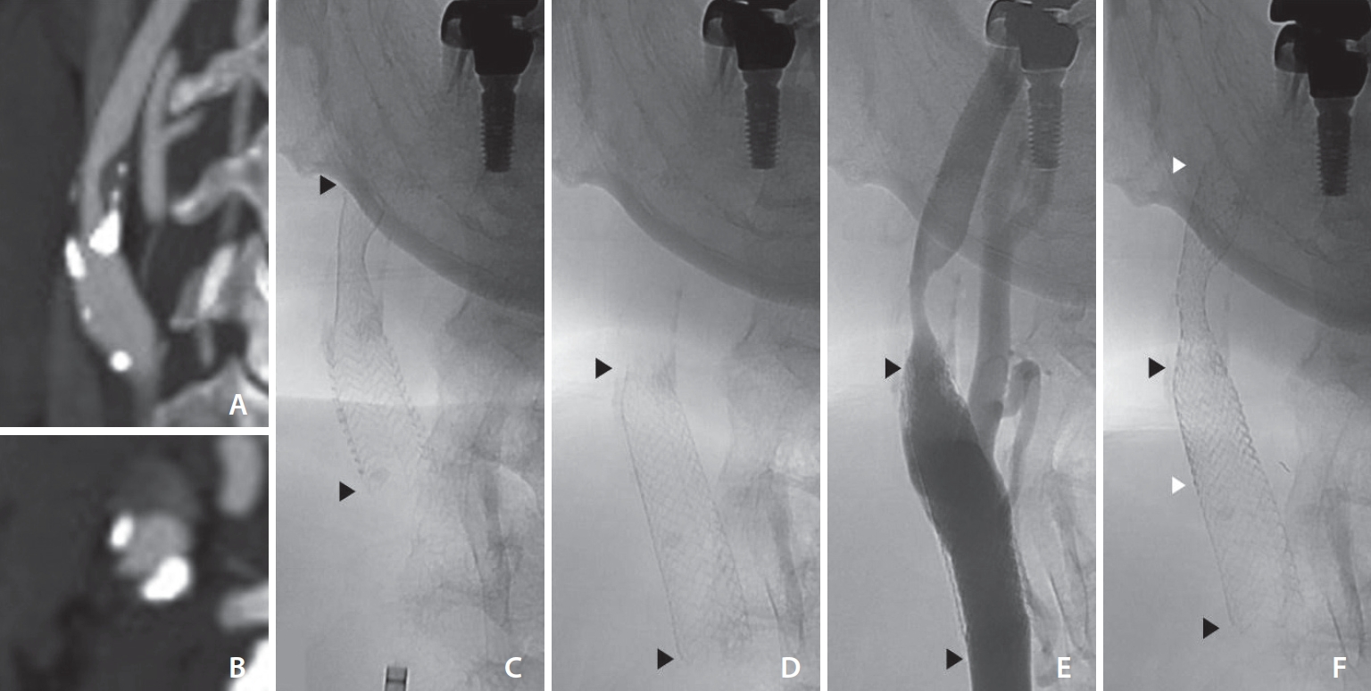
A 65-year-old male presented to the hospital with intermittent left arm weakness. The patient had a medical history of tobacco use, coronary artery disease, dyslipidemia, and hypothyroidism. He was on medical treatment with daily doses of aspirin 100 mg, atorvastatin 10 mg, ezetimibe 10 mg, and levothyroxine 0.1 mg. Brain computed tomography angiography (CTA) revealed a significant stenosis of the right proximal internal carotid artery (ICA) (Fig. 1A, B) and magnetic resonance imaging (MRI) showed perfusion delay in the relevant arterial territory without evidence of acute cerebral infarction. Because of his recurrent ischemic symptoms, carotid stenting was planned and clopidogrel 75 mg daily was started 10 days before admission.
Under local anesthesia with 1% lidocaine via the right common femoral artery, a 90 cm long 8F Fubuki guiding catheter (Asahi Intec) was placed in the right common carotid artery. The stenosis of the right proximal ICA was measured to be 15 mm in length with 60% stenosis according to the North American Symptomatic Carotid Endarterectomy Trial (NASCET) criteria. The gradient of vessel diameter was not large with a proximal diameter of 8.4 mm and a distal diameter of 6.1 mm. After the placement of the Emboshield NAV6 (Abbott Vascular), an Acculink RX tapered stent (7–10×40 mm; Abbott Vascular) was deployed across the lesion with sufficient proximal and distal margins (Fig. 1C). Poststent balloon angioplasty was not performed due to adequate stent lumen. Control angiography showed patent stented segment of the right ICA with adequate stent apposition to the vessel wall. The patient was discharged home 2 days later with no neurological deficits and continued dual antiplatelet therapy.
Four months after discharge, the patient presented to our hospital with a complaint of right facial (parotid area) pain, tenderness and swelling for 5 days. Neck CT with contrast suggested parotitis and downward migration of the stent. After a few days, the patient complained a foreign body sensation in the right neck. Follow-up brain CTA and MRI showed downward migration of the stent with significant stenosis at the previous lesion of the right ICA stenosis and perfusion delay in the relevant arterial territory.
One week after the follow-up MRI, the patient was readmitted for management of carotid restenosis. The right carotid angiogram confirmed stent migration and restenosis of the previous stenotic lesion with 70% stenosis (Fig. 1D, E). Compared to the previous angiogram, the stent had migrated approximately 14 mm inferiorly. Pre-stenting balloon angioplasty was performed with a 4 mm×40 mm Aviator Plus balloon (Cordis). An 8 mm×40 mm Precise RX stent (Cordis) was successfully deployed across the stenotic lesion with the Emboshield NAV6. The stent was deployed overlapping the distal half of the previously delivered stent (Fig. 1F). The control angiogram showed a spanned stenotic carotid segment with improved antegrade flow in the right ICA. The patient was discharged home 2 days after the procedure with no neurological complications. Since then, the patient did not complain of any signs or symptoms, and no further studies were performed.

DISCUSSION

Stent migration after CAS is a rare complication but has been reported to occur infrequently depending on the patient’s lesion and associated carotid arteries [8,9]. Excessive vessel tortuosity or the formation of dense fibrosis after CEA may increase the risk of stent migration. Some authors reported that stent migration was observed when the lesion had severe stenosis with excessive kinking of >90 degrees at the distal end or when the lesion had a previous CEA [10,11]. However, in our case, the stenotic vessel had a calcified lesion without tortuosity, and an open-cell stent was used with a length and diameter sufficient to cover the stenotic lesion and approximate the normal arterial wall.
Interestingly, stent migration does not appear to be affected by stent design, such as open-cell [7,10] or closed-cell [4,6]. Closed-cell stent migration may result from the watermelon-seeding effect, where the stent moves toward the larger proximal vessel due to diameter differences [6]. In contrast, open-cell stents offer flexibility and resistance to deformation, allowing independent segment expansion even in vessels with diameter variations. In addition, open-cell nitinol stents are generally less prone to migration. In this case, the diameter difference between the two ends of the vessel was only 2.3 mm, and despite the use of an oversized tapered open-cell nitinol stent, stent migration occurred.
In published cases, stent migration was observed not only during follow-up, as in our case, but also immediately after CAS (Table 1) [4-7,10]. In general, endothelial growth after stenting integrates the stent into the vessel wall and prevents migration. Within a few weeks, the stent struts are expected to be integrated into the vessel wall, making migration or dislodgement difficult. Therefore, stent migration, if it occurs, is likely to have occurred within a few days after CAS.
In summary, we have demonstrated the rare case of delayed migration of a tapered open-cell nitinol stent in the absence of risk factors for stent migration. Clinicians should be aware of this rare complication after CAS and early follow-up with noninvasive imaging should be considered if the lesion is at risk for stent migration.

Notes

Fund

None.

Ethics Statement

This study was reviewed and approved by Yonsei University Gangnam Severance Hospital, Institutional Review Board (3- 2022-0195), and the patient provided written informed consent for the publication of the case report.

Conflicts of Interest

SHS has been the Editor-in-Chief of the Neurointervention since 2023; however, SHS has not been involved in the peer reviewer selection, evaluation, or decision process of this article. No potential conflict of interest relevant to this article was reported.

Author Contributions

Concept and design: IHL, TO, and SHS. Analysis and interpretation: IHL, TO, and SHS. Data collection: IHL, TO, and SHS. Writing the article: IHL, TO, and SHS. Critical revision of the article: IHL, TO, YHJ, KYL, and SHS. Final approval of the article: IHL and SHS. Overall responsibility: SHS.

Fig. 1.
(A, B) Brain computed tomography angiography showing severe stenosis with calcified plaque. Fluoroscopic images showing (C) the initial stent insertion, (D) downward migration of the stent on 4 months follow-up, (E) restenosis distal to the stent, and (F) after the second stent insertion. Arrowheads point to both ends of the initial stent (black) and the second stent (white).
neuroint-2023-00304f1.jpg
Table 1.
Summary of case reports for the stent migration
Ref No. Authors (y) Sex/age Carotid artery stent
Stent migration
Retreatment Follow-up
Indication Stent Duration/study f/u Finding Related factor
4 Castriota et al. 2008 M/69 Severe (95%) asymptomatic, right. SES, overlapped by Wallstent (nontapered closed-cell) 15 mo/CDU Distal migration Tortuosity distal to stenosis. 7.0×30 mm Carotid Wallstent Stable on 8 mo, 10 mo, and 13 mo CDU
5 Yan et al. 2009 M/5 Williams syndrome. BES (Genesis), nontapered 7 mo/DSA Distal migration of BES to the distal CCA None Stable for 5 y
Symptomatic stenosis in right brachiocephalic artery (90%) and left CCA (60%).
6 Badruddin et al. 2011 M/70 Severe symptomatic (TIA) stenosis (99%), left. SES, Xact, 7×20 mm, nontapered closed-cell 8 mo/CTA Proximal downward migration Relatively small sized and short stent. Tapered 6–8×40 mm Xact stent f/u
Calcified stenotic lesion.
7 Endo et al. 2023 M/70 Restenosis (80%) 6 months after CEA for symptomatic right ICA stenosis (86%) due to radiotherapy. SES, tapered open-cell stent (Protégé RX 10/7×40 mm) #2 d/CTA Proximal migration into CCA Large difference between proximal (7.4 mm) and distal (2.8 mm) vessel diameters. None Stable on 6 mo DSA f/u
Post-RTx and endarterectomy (patch graft) state. Stable for 3 y
10 Swarnkar et al. 2014 M/63 Restenosis after CEA, bilaterally. SES, nontapered, overlapping stents Right: #4 d/DSA. Right: caudal migration of proximal stent and cranial migration of distal stent (9 mm gap) Bilateral carotid CEA complicated by hematoma formation. Right: during procedure; additional stent (Acculink 6×20 mm) #5 d
Right: 90% stenosis with 5.2 cm in length. Right: Acculink 5×40 mm and 5×20 mm Left: #2 d/CDU Fibrosis and loss of elasticity at the surgical site. Right: stable
Left: 80% stenosis with 7 cm in length. Left (#4 days after right): Acculink 5×40 mm and 6×40 mm Left: 12 mm separation #4 days after stent: 2nd additional stent (Acculink 6×40 mm) Left: observation for migration

Ref, reference; f/u, follow-up; M, male; SES, self-expandable stent; CDU, color doppler ultrasound; CCA, common carotid artery; BES, balloon expandable stent; DSA, digital subtraction angiography; TIA, transient ischemic attack; CTA, computed tomography angiography; CEA, carotid endarterectomy; ICA, internal carotid artery; RTx, radiotherapy.

REFERENCES

1. Baek JH. Carotid artery stenting for asymptomatic carotid stenosis: what we need to know for treatment decision. Neurointervention 2023;18:9-22.
crossref pmid pmc pdf
2. Hill MD, Brooks W, Mackey A, Clark WM, Meschia JF, Morrish WF, et al, CREST Investigators. Stroke after carotid stenting and endarterectomy in the Carotid Revascularization Endarterectomy versus Stenting Trial (CREST). Circulation 2012;126:3054-3061.
crossref pmid pmc
3. Seo KD, Lee KY, Kim BM, Suh SH. Delayed cerebral infarction due to stent folding deformation following carotid artery stenting. Korean J Radiol 2014;15:858-861.
crossref pmid pmc
4. Castriota F, Martins EC, Dall’Olmo CA, Molnar RG, Manetti R, Liso A, et al. Stent migration as a late complication following carotid angioplasty and stenting. EuroIntervention 2008;4:397-399 discussion 400-404
crossref pmid
5. Yan BP, Rosenfield K, Garasic JM, de Moor M. Carotid artery stent migration in a child with Williams syndrome. Pediatr Cardiol 2009;30:551-552.
crossref pmid pdf
6. Badruddin A, Lazzaro MA, Taqi MA, Zaidat OO. Downward migration of carotid stent on 8 months follow-up imaging: possible stent “watermelon- seeding” effect. J Neuroimaging 2011;21:395-398.
crossref pmid
7. Endo H, Ishizuka T, Murahashi T, Oka K, Nakamura H. Proximal migration of a tapered open-cell stent after carotid artery stenting for restenosis following endarterectomy. J Stroke Cerebrovasc Dis 2023;32:106953
crossref pmid
8. Kokkosis AA, Macdonald S, Jim J, Shah R, Schneider PA. Assessing the suitability of the carotid bifurcation for stenting: anatomic and morphologic considerations. J Vasc Surg 2021;74:2087-2095.
crossref pmid
9. Kumins NH, King AH, Ambani RN, Thomas JP, Kim AH, Augustin G, et al. Anatomic criteria in the selection of treatment modality for atherosclerotic carotid artery disease. J Vasc Surg 2020;72:1395-1404.
crossref pmid
10. Swarnkar A, Krishna SH, Zimmerman N, Latorre J, Deshaies EM. Bilateral migration of open-cell design carotid stents in the early post-procedure period: a rare complication. Interv Neuroradiol 2014;20:761-765.
crossref pmid pmc pdf
11. Roubin GS, Iyer S, Halkin A, Vitek J, Brennan C. Realizing the potential of carotid artery stenting: proposed paradigms for patient selection and procedural technique. Circulation 2006;113:2021-2030.
crossref pmid


ABOUT
ARTICLE CATEGORY

Browse all articles >

BROWSE ARTICLES
AUTHOR INFORMATION
Editorial Office
#705-2C, 9 Gangnam-ro, Giheung-gu, Yongin-si, Gyeonggi-do 16977, KOREA
Tel: +82-31-994-4382    Fax: +82-2-2275-9198    E-mail: editor@neurointervention.org                

Copyright © 2024 by Korean Society of Interventional Neuroradiology.

Developed in M2PI

Close layer
prev next