INTRODUCTION
Cerebrovascular disease is one of the leading causes of death in Korea, and interventional neuroradiology (INR) procedures have increased substantially in both number and complexity. As the volume and types of procedures grow, we will start to see lengthy procedures to achieve better results. However, interventional radiological procedures can potentially expose a patient to high doses of radiation, which requires particular concern and continuous monitoring [1,2].
Since interventional radiology involves prolonged use of X-rays, reference levels (RLs) for various procedures might be the next step in quality assurance and improvement [3]. Similar to diagnostic reference levels (DRLs), continuously updated RLs in diagnostic and interventional radiology should provide a framework for physicians performing endovascular treatment in order to control, limit, and reduce radiation exposure for patients and personnel [3-5].
Previous studies in Korea and other countries have investigated RLs and reported large variation within hospitals using different RL guidelines [5-10]. Recognizing the need for continuous dose monitoring in diagnostic cerebral angiography and the fact that no such multi-center study has ever been carried out regarding other INR procedures, the authors collected data concerning the dose to patients undergoing some of the most common INR procedures performed in Korea.
The purposes of the present study were (1) to obtain baseline radiation dose data by evaluation of patient exposure at multiple centers, (2) to compare these data with the recent literature from other studies, and (3) to propose new DRLs.
MATERIALS AND METHODS
Study Design and Patient Selection
This was a multicenter retrospective study with the participation of 22 hospitals involving prospectively and consecutively collected data between December 2020 and June 2021. The target hospitals were secondary or tertiary hospitals designated as local medical centers in South Korea and endovascular treatment-capable institutions. As radiation doses show a wide distribution with respect to pathology and type of neurointerventional procedure, we focused on 4 standardized cerebral angiographic examinations. Each center was asked to register 20 cases of diagnostic cerebral angiography, 15 cases of aneurysm embolization, 15 cases of stroke thrombolysis/thrombectomy, and 5 cases of arteriovenous malformation (AVM) embolization, respectively. In diagnostic angiography, the age of participants ranged from 17 to 86 years with mean age of 56 years. For each examination, the centers were asked to fill out a questionnaire containing various information regarding radiation data. The X-ray systems used in this study were all biplane digital subtraction angiography (DSA) (14 Siemens, 7 Philips, and 1 GE machines). Institutional ethics board approval was granted for this retrospective descriptive cohort study performed at various institutions. A waiver of the need for consent was obtained for this Health Insurance Portability and Accountability Act compliant survey research.
Procedures Included
The neurointerventional procedures were divided into 2 groups: (1) diagnostic cerebral angiography for aneurysm evaluation and (2) therapeutic procedures, namely aneurysmal coil embolization, mechanical thrombolysis/thrombectomy for ischemic stroke, and AVM embolization. For a diagnostic angiography procedure, we focused on aneurysmal evaluation, and follow-up after clipping or coiling of an aneurysm was excluded. The procedures were performed by an experienced interventional neuroradiologist or clinical fellow undergoing interventional neuroradiology training, all using their own protocols.
Data Collection
Collected patient radiation dose indicators were as follows: dose–area product (DAP), cumulative air kerma (CAK), fluoroscopic exposure time, and number of angiographic image acquisitions. Collected data were entered onto a Microsoft Office Excel 2010 (Microsoft, Redmond, WA, USA) spreadsheet. Patient sex, patient age, procedure type, and number of exposures were recorded for all procedures. For the aneurysm coiling procedure, the following additional parameters were recorded: location, size of aneurysm, embolization technique, and presence of peri-procedural complication. For stroke thrombolysis, the following additional parameters were recorded: level of occlusion, thrombectomy technique (stent retriever, direct aspiration, or combined), trial number of device passage, and final angiographic results. For AVM embolization, the following additional parameters were recorded: size and location of AVM nidus, Spetzler–Martin grade, and purpose of embolization. Data were analyzed to assess mean±standard deviation for each parameter. As collected radiation doses will show a skewed distribution with extreme values and a long upper tail, 75th percentile values were also analyzed to propose DRL.
Statistics/Data Analysis
A descriptive analysis of the data was performed. Statistical analysis was performed with Statistical Package for Social Scientists (SPSS) software version 23 (IBM, Armonk, NY, USA). Median, mean, and maximum and minimum radiation doses were calculated for each of the 4 procedures. Except for diagnostic angiography, other procedure-related variables were analyzed using 2 different statistical tests depending on the number of groups per variable. Variables were compared according to the t-test if data were normally distributed between 2 groups. Between 3 groups, when statistically significant differences occurred, the ANOVA test with Tukey’s hostly significant difference (HSD) post hoc test for multiple comparisons was performed. A P-value of less than 0.05 was considered statistically significant.
RESULTS
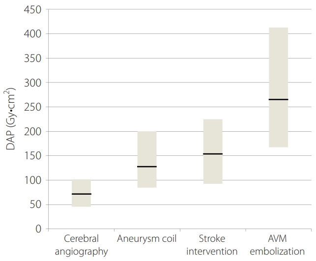
A total of 1,160 patients who underwent neurointerventional procedures were eligible for inclusion. Of the 1,160 procedures, 429 diagnostic cerebral angiograms, 327 aneurysm coilings, 326 stroke thrombolysis, and 78 AVM embolization procedures were performed. Median DAPs were as follows: diagnostic cerebral angiography, 70.4 Gy·cm2; aneurysm coiling, 130.6 Gy·cm2; stroke thrombolysis 150.4 Gy·cm2; and AVM embolization, 264.3 Gy·cm2. Detailed DAP distributions are illustrated in Table 1 and Fig. 1 demonstrates the range of radiation doses for each procedure type.
In diagnostic cerebral angiography, the mean DAP±standard deviation was 78.0±43 Gy·cm2 for DAP, 541.5±333.2 mGy for CAK, 10.4±6.4 minutes for fluoroscopic time (FT), and 511.1±208.5 frames for angiographic image frames. The third quartiles, which may be set as a DRL, were 101.6 Gy·cm2 for DAP, and 711.3 mGy for cumulative air kerma, 13.3 minutes for fluoroscopy times, and 637 for number of image frames.
Radiation Dose for Aneurysm Coiling
Patient characteristics are summarized in Table 2. We identified a total of 327 patients (30.2%; 99 males, and 69.8%, 228 females, mean age 60.9 years) with either an unruptured or ruptured intracranial aneurysm. The median aneurysm size was 5.4 mm, with a minimum diameter of 2.3 mm and a maximum diameter of 18.1 mm. In total, 279 (85.3%) out of a total of 327 aneurysms were located at the anterior circulation and 48 (14.7%) aneurysms at the posterior circulation. Regarding the endovascular treatment technique, 159/327 (48.6%) patients were treated with a simple or double microcatheter technique, 157/327 (48.0%) by a balloon or stent-assisted technique, and 11/327 (3.4%) by using a flow diverter. Of the 327 patients, we had 14 (4.3%) cases of peri-procedural complications. Results of radiation doses and fluoroscopy time are illustrated in Tables 1 and 3. In aneurysmal embolization, the mean DAP±standard deviation was 151.0±96.8 Gy·cm2 for DAP, 2,622.8±2,110.7 mGy for CAK, 45.8±30.7 minutes for FT, and 803.7±540.0 frames for angiographic image frames. The third quartiles, which may be set as a DRL, were 199.9 Gy·cm2 for DAP, and 3458.7 mGy for cumulative air kerma, 57.3 minutes for fluoroscopy times, and 1,000 for number of image frames.
Pairwise comparison of mean DAP between different sex groups reached statistical significance (P<0.05; Table 3). Concerning aneurysm location, mean DAP, air-kerma (AK), and FT values did not show statistical significant difference (P>0.05; Table 3). Concerning the presence or absence of peri-procedural complications, pairwise comparisons of mean DAP, AK, and FT groups were significantly different (P<0.05; Table 3). The mean DAP of the simple (or double) microcatheter technique was 143.3±94.88 Gy·cm2, 160.7±99.7 Gy·cm2 for assisted (balloon or stent) technique, and 125.5±72.1 mGy for flow diverter. Pairwise comparisons of mean DAP between the 3 groups did not reach statistical significance.
Radiation Dose for Stroke Thrombolysis
Patient characteristics are summarized in Table 4. A total of 326 patients received mechanical thrombectomy, with 140 (42.9%) males and 186 (57.1%) females. The mean age was 70.8 years old. The median NIHSS was 13, range between 2 and 40. Out of the 326 thrombectomized patients, 283 (86.8%) cases developed in the anterior circulation and 43 (13.2%) in the posterior circulation. Regarding the thrombolysis technique, 80/326 (24.5%) patients were treated by an aspiration technique, 113/326 (34.7%) by a stentriver, and 133/326 (40.8%) by a combination technique. Of the 326 patients, we had 278 (85.3%) cases of successful recanalization (greater than thrombolysis in cerebral infarction scale grade 2b). The trial number of device passage for thrombectomy were as follows: once (139 cases, 42%), twice (77 cases, 24%), and more than 3 times (110 cases, 34%), respectively.
Results of radiation doses and fluoroscopy times are illustrated in Tables 1 and 5. In stroke thrombolysis, the mean DAP±standard deviation was 176.2±118.7 Gy·cm2 for DAP, 1,263.5±918.2 mGy for CAK, 35.1±26.4 minutes for FT, and 630.2±619.0 frames for angiographic image frames. The third quartiles, which may be set as a DRL, were 225.1 Gy·cm2 for DAP, 1,590 mGy for cumulative air kerma, 44.7 minutes for fluoroscopy times, and 800 for number of image frames.
Pairwise comparison of mean DAP, AK between different sex groups reached statistical significance (P<0.05; Table 5). Concerning the occlusion site and result of successful recanalization, mean DAP, AK, and FT values did not show statistical significant difference (P>0.05; Table 5). Concerning the trial number of device passage, pairwise comparison of mean DAP, AK, and FT between groups were significantly different for all variables (P<0.05; Table 5). Pairwise comparision of different thrombolysis technique groups reached statistical significance in terms of mean DAP, AK, and FT, especially between aspiration and combined technique or between stentriever and combined technique groups.
Radiation Dose for AVM Embolization
Patient characteristics are summarized in Table 6. A total of 78 AVM cases were treated, 42 (53%) were male and 36 (47%) were female. The mean age was 40.6 years old. The median size of AVM nidus was 2.5 cm, ranging from 0.8 cm to 6.1 cm. Out of the 78 patients, 59 (75.6%) AVM procedures were performed at the anterior circulation and 19 (24.4%) at the posterior circulation. Regarding the purpose of embolization of AVM, 22 (28.2%) patients were treated before operative resection, 21 (26.9%) patients before or after radiosurgery, and 35 (44.9%) patients for complete cure. Spetzler–Martin grades of pre-embolization were I in 18 cases (23%), II in 36 (46 %), III in 18 (23%), IV in 5 (6%), and V in 1 (1%), respectively.
Results of radiation doses and fluoroscopy time are illustrated in Tables 1 and 7. In AVM embolization, the mean DAP±standard deviation was 300.1±184.2 Gy·cm2 for DAP, 3,673.8±2,923.0 mGy for CAK, 77.2±50.1 minutes for FT, and 1,293.8±1,061.6 frames for angiographic image frames. The third quartiles, which may be set as a DRL, were 412.3 Gy·cm2 for DAP, 4,447.8 mGy for cumulative air kerma, 99.3 minutes for fluoroscopy times, and 1,621.3 for number of image frames.
Pairwise comparison of mean DAP and AK between different sex groups did not reach statistical significance (P>0.05). Concerning the AVM location, mean DAP, AK, and FT values did not show statistical significant difference (P>0.05). Concerning the size of the nidus, pairwise comparison of mean DAP, AK, and FT between groups did not reach statistical significance (P>0.05). Concerning the purpose of embolization, pairwise comparison between groups revealed statistical significance in terms of mean DAP, AK, and FT. Radiation dose tended to increase in the curative embolization group.
DISCUSSION
Neurointerventional procedures are increasingly used. Due to the complexities of the cerebrovascular anatomy and the procedures themselves, these procedures often require a long time to perform. With more and more complex procedures, radiation dose for patients is an important issue. As a tool for optimization and quality improvement of practices, the need for establishing and monitoring radiation doses for neurointerventional procedures is obvious.
According to the ICRP 135 publication [4], application of several radiation dose metrics (e.g., DAP and FT) is recommended for DRL establishment of fluoroscopically guided interventions. In this context, the DRL value is defined as the 75th percentile of the distribution of the DRL quantity, representing a commonly calculated radiation dose metric in neurointerventional procedures.
Our data showed great variability of dose levels between categories, up to a 3.8-fold difference in median values between diagnostic cerebral angiography and AVM embolization, mainly due to the difference in procedure complexity and operator and institutional experience.
Table 8 shows a comparison between our results and those published in the literature [8-14]. For cerebral angiography, when comparing radiation dose with that in the available literature, DAP, AK, and FT were found to be comparable. While comparing the radiation dose with our previous data [8], third-quartile DAP and AK values were found to be significantly lower than the published reference (Table 8). A significant dose reduction in 5 out of 22 hospitals was attributed to the introduction of an advanced technology system. The dose reduction in the other 8 centers was achieved by privileging a low–dose technique (low dose fluoroscopy and reducing the number images in DSA).
For aneurysmal embolization, when comparing radiation dose with that in the available literature, DAP, AK, and FT were found to be comparable. However, when comparing radiation dose with our previous data, third-quartile DAP and AK values were found to be significantly lower than the published references (Table 8). While fluoroscopy time was comparable to other studies, the number of angiographic images was much higher.
In the present study, DAP of aneurysm treatment was not significantly different in terms of endovascular technique and aneurysmal location, but was associated with sex and procedural complications. Application of a stent or balloon-assisted technique in more complex aneurysms yielded a higher AK (mean 2,994 mGy) and FT (mean 51 minutes) when compared with a simple catheter technique using 1 or 2 microcatheters (P<0.05). When we assessed the procedural factor contributing to increased radiation dose during aneurysmal coiling, the occurrence of complications resulted in increased patient dose, as additional imaging was required to treat the complications. However, the location of an aneurysm was not a significant factor for increased radiation dose. Acton et al. [15] argued that aneurysm location is the biggest determinant of radiation dose during coiling procedures; therefore, they suggested separate RLs between anterior and posterior circulation coiling procedures. As wide variation in the RLs for intracranial aneurysmal embolization was evident, the results of this study highlight the need to monitor doses for aneurysm coiling procedures in each country.
For stroke intervention, to our knowledge, this study provides the first radiation data related to mechanical thrombolysis in Korea. When comparing radiation dose with recent other studies, all radiologic dose parameters for our data were found to be higher than in other studies [13,14].
In the present study, DAP was not significantly different in terms of occlusion site, result of successful recanalization, but higher radiation dose was associated with sex, number of device passages, and occlusion removal technique. Male patients received higher doses than female patients, although sex was not suspected to be a factor influencing the radiation dose in stroke interventions. However, we did not consider the patient’s morphology, as this might be a confounding factor, considering that men are generally heavier than women. As we usually use an automatic exposure control system for the fluoroscopic machine in most of our procedures, different habitus can affect the dose production setting to maintain image brightness controlled by automatic exposure algorithms. While the number of attempts required to remove a thrombus is known to reflect the complexity of the procedure, this parameter proved to be the most significant factor for affecting patient dose. Although radiation dose for a combined technique was higher than for a single device technique, no significant difference in dose between aspiration and stent retriever techniques was found. These results suggest either aspiration or stent retriever can be used if there is no difference regarding clinical outcome for dosimetric considerations.
The establishment of useful local RLs also requires the inclusion of sufficient data, which can be a challenge for individual neurointerventional units. This was observed in the setting of AVM treatment in the present study. The number of AVM procedures performed was relatively small, and the 75th percentile DAP value was 412 Gy·cm2. When comparing radiation dose with the available literature, a large variability was evident [12]. Our results were generally comparable to other reported data.
Setting up DRLs for dose-intensive examinations involving fluoroscopy is a difficult task due to the large variability in FT and the number of images, leading to a wide distribution of patient doses. Furthermore, unique institutional patterns and operator preferences for INR procedures can cause difficulty in making radiation dose comparisons between hospitals [16]. Nevertheless, this study indicates our values of various radiation doses of INR procedures were within the range of previous published reports and concordance of the data with those reports. Therefore, our proposed RLs would be valuable in comparing and monitoring radiation doses.
INR procedures show wide variety and complexity, and are continuously progressing, so radiation dose may be higher with complex, newer, or meticulous procedures [17]. Nevertheless, it is each practitioner’s responsibility to investigate his or her own practice and to limit unnecessary radiation exposure according to the ALARA (As Low as Reasonably Achievable) principle [18-21].
This study has limitations. First, we collected retrospective registry data even though we designed a prospective study without controlling for potential sources of bias. Second, our data did not include information on the patient’s body mass index or factors that determine the complexity of various procedures. However, previous studies have shown that the amount of radiation for INR procedures is much more affected by procedure complexity than by patient size and weight. Miller et al. [12] considered that it is sufficient to use reference levels that have not been corrected for patient body habitus. Third, we did not evaluate the complexity of the various neurointerventional procedures. Further investigation with larger populations and prospective evaluation is clearly warranted in order to validate our results.
CONCLUSION
In this study, patient radiation exposure was collected and analyzed for various neurointerventional procedures of varying complexities and found to be comparable to the published literature. Various dose reference levels introduced in this study promote the optimization of patient doses in diagnostic and therapeutic INR procedures. Proposed third quartile DAP (Gy·cm2) values were 101.6 for diagnostic cerebral angiography, 199.9 for aneurysm coiling, 225.1 for stroke thrombolysis, and 412.3 for AVM embolization. Continual evolution of practices and technologies requires regular updates of reference levels.